Evelyn Witkin’s Lasker Award

Dr. Witkin in Plos Genetics  2012.

Dr. Witkin in Plos Genetics 2012.

I was thrilled to hear the news that Dr. Evelyn Witkin won this year’s Lasker Award for her pioneering work in DNA damage response. Witkin started out as a biology student at NYU; originally planning to continue on at NYU for her graduate work she was “bounced out of college” her senior year for her role in protesting NYU athletics’ acquiescence to segregation in the South. Fortunately, her move to Columbia and then Cold Spring Harbor Laboratory was a turning point in her career (mind you, NYU didn’t even have a genetics department).

At CSHL, Witkin discovered a strain of bacteria that was immune to the damaging effects of UV radiation. This was the starting point for understanding the molecular pathways that regulate DNA damage and repair (which so often go haywire in cancer).

I first contacted Witkin in 2013, while I was trying to dig up information about another CSHL alum, Martha Chase. While Martha remained elusive, I got a broader sense of what it was like to be a female scientist at CSHL in the 40s and 50s (surprisingly good by her account). On top of being a top-notch scientist, it was evident from my correspondence with her that Witkin is a remarkably kind, I’d say graceful, person. I’ve included an email from her below to give you an idea.

Hello Kristen,

I remember Martha Chase very well, as an attractive young woman who worked with Al Hershey, but I’m afraid I will have to disappoint you when I tell you that I really did not know her very well.   I don’t recall ever talking with her about the work she was doing with Al, either before or after the publication of their 1952 paper. We exchanged smiles and greetings when our paths crossed, which somehow did not happen very often.  Perhaps that was because by 1952, I was no longer living on the grounds at the lab, but across the harbor in CSH village.

I appreciate your interest in what CSH was like in the forties and fifties.  I will try to answer your specific questions as soon as I can, but it  occurred to me that you might want to read my memoir Chances and Choices: Cold Spring Harbor 1944-1955, which was published in 2002 in the Annual Review of Microbiology.  I have attached it here.  Perhaps you will find some of your questions answered there.  After you have had a chance to read it, it might be useful for you to send me an amended list of questions, which I will then be glad to answer as best I can.  

I have to call your attention to an error in that article, which I discovered only after it was published.  In the list of future Nobel laureates that I met at CSH.  I mistakenly included O.T. Avery,  C. M. McCleod and M. McCarty.  I has assumed that the prize had been awarded to them, and was shocked to learn, that it had not.

I have always felt that i was amazingly fortunate to have been at the CSH Lab as a graduate student, postdoc and staff member, during those indescribable tears of ferment and promise.  I made my first visit there in 1943, which, if my arithmetic is right, is now seventy. years ago.  Yet I have vivid memories of so much that happened there, and of so many of the people who helped make molecular biology happen.  There was a shared excitement that made every day an adventurea   

I’m sure that you must be having a wonderful experience in your graduate work in 2013.  Whenever I visit,  I sense that a lot of the magic that I remember is still there, on steroids.   

Thank you for writing, Kristen.  Let’s keep in touch.

All best wishes, Evelyn

CSHL Symposium Haikus

Monday and Tuesday marked the 26th annual Cold Spring Harbor In-House Symposium when the lab community comes together to hear research updates from half of the faculty. As corny as it sounds, I think that the couple days before leaving to spend Thanksgiving with family is the perfect time to celebrate the scientific progress of our colleagues. During last night’s banquet, as we gathered around to hear David Spector’s Symposium top ten list, there was a palpable sense of gratitude. It’s a tradition I’ll miss when I leave CSHL.

Anyway, I was the chair for Tuesday’s morning session, and had to scramble for something creative to do for my introductions. Swinging away from the previous day’s “roast” I decided (for some reason) to do haikus. Here they are for the speakers; you can click on the photos to read about each lab’s research.

Tom Gingeras

Across vast stretches
The genome yields not protein
What does it ENCODE?

Justin Kinney

More than location
The promoter sequence charts
The cost of binding

Grisha Enikolopov

In the zone they wait
Until spark disrupts slumber
A neuron is born

Adrian Krainer (avid volleyball player)

Bump, set, spliceosome
Better to skip than be skipped
Whose alternative?

Ivan Iossifov

The truth is out there
Unlock the text, give structure
From many labs, one

Leemor Joshua-Tor

Diffracting crystals
Bare structures, mechanisms
In purple-walled rooms
-Eugene Plavskin

Particles and Waves
Interfere, Diffract, and Give
Crystal clear answers
-Jonathan Ipsaro

Here are some more haikus from Symposium attendees:

What is sarin?

UN chemical weapons experts view victims of last week’s attack near Damascus, Syria photo credit: Abo Alnour Alhaji/Reuters

At around 2 a.m. on Wednesday August 21st, a barrage of rockets exploded “like water tanks bursting” and released chemicals into two neighboring suburbs of Damascus. Still sleeping civilians inhaled these chemicals, and many never made it out of their beds. As reported by the New York Times, those who did arrived at hospitals, with “their bodies convulsing and mouths foaming. Their vision was blurry and many could not breathe.”

Hamish de Bretton-Gordon, the former commander of the chemical, biological and nuclear counter-terrorism unit at Britain’s defense ministry was quoted as saying, “If it is a chemical weapon, it’s most likely to be a nerve agent – and we know that sarin has been used in the past in Syria. We know that Assad has very large stockpiles of sarin, and a delivery of sarin would create these kinds of casualties.”

So what is the nerve gas, sarin, and what makes it so deadly?

Space-filling model and line structure of sarin.

Space-filling model and line structure of sarin.

Sarin is an organic compound that contains a carbon-phosophorous bond. Compounds like it are often used as insecticides and are extremely potent blockers of communication between nerves and organs.

Sarin, and other nerve agents implicated in the Damascus attacks (tabun, soman, and VX) block communication between nerves and organs by allowing the build up of a neurotransmitter called acetylcholine.

Normally, an enzyme called acetylcholinesterase rapidly breaks down acetylcholine after it’s been released from a nerve ending. At the junction between nerve and muscle acetylcholinesterase works as a sort of “on-off switch” for muscle contraction. When acetylcholine is released from a nerve cell it binds to receptors on muscle and makes it contract. Normally, acetylcholine molecules are kicked off of the muscle receptors and broken down by acetylcholinesterase, allowing the muscle to relax until the next incoming nerve impulse releases acetylcholine.

Sarin breaks this on-off switch by attaching itself to acetylcholinesterase, blocking its ability to break down acetylcholine. This allows it to build up in the space between the nerve and muscle and continuously stimulate muscle, leading to spasms and eventual paralysis. The same process simultaneously takes place at nerve-nerve cell junctions throughout the body: salivary glands are overactivated, causing hyper salivation and mouth-foaming; pupils constrict, impairing vision; and bronchial tubes squeeze shut, suffocating the victim. Sarin is devastatingly effective, and its symptoms line up with those reported for hundreds* of people in the Damascus suburbs.

For a short while, before supplies ran out, the victims were administered an antidote to sarin, called atropine. Atropine acts by competing with acetylcholine for binding to receptors on the muscle (or other nerve cell), while not activating the muscle or nerve cell itself. This gives the muscles and nerve cells relief from stimulation.

Atropine blocks acetylcholine from binding to muscle or other nerve cell receptors. Acetycholinesterase enzyme, shown right, is inactivated by sarin binding.

Atropine blocks acetylcholine from binding to muscle or other nerve cell receptors. Acetycholinesterase enzyme, shown right, is inactivated by sarin binding.

Unfortunately even if atropine is administered, sarin – particularly its effects on breathing – can have longterm negative consequences on brain function. There are fears that current atropine supplies are very limited, and that any existing stockpiles are likely controlled by the Syrian military.

Secretary of State of John Kerry has condemned the attacks as a “moral obscenity“, and the Los Angeles Times reports that Kerry’s emotional language may be an attempt to “build up public support for a muscular attack” on Assad’s forces.

Sources:

Los Angeles Times 8/26/13

New York Times 8/26/13

Reuters 8/23/13

Chiral publishing group “Physiological Effects of Nerve Gas”

corrected: Wednesday, not Monday August 21st

*We now know the attack likely affected thousands, with the death toll at 1,429 as of 8/31

Updates:

NYTimes reports death toll was so high because rockets delivered large payloads of sarin gas.

Re-turning the favor

Since I can remember, summers for me meant the shore. When my siblings and I weren’t playing leap frog with the waves or making drizzle sand towers, we patrolled the beach. We buried the jellyfish that washed up on shore, lest anyone fall victim to an ill-placed foot. We picked up stray trash so that it wouldn’t hurt animals like those 6-pack rings did. My favorite duty, however, was gingerly righting the horseshoe crabs that had the misfortune of winding up on their backs.

Looking like a primordial holdover, the horseshoe crab easily evoked my sympathies. It reminded me of The Little House: sturdily built to last the ages, it was made to look lumbering by the civilization that sprung up around it. And to think 300 million years ago it was the dominant creature on the planet. Further proof they get no respect: they aren’t even crabs! The horseshoe crab is an arthropod that’s more closely related to spiders or scorpions than any of the crustaceans.

From The Little House by Virginia Lee Burton

My grandma’s house sits on the southern tip of New Jersey on the Delaware Bay, which happens to be prime breeding ground for the horseshoe crab. Every year as my family descended on the beach on the bay, the horseshoe crabs came to mate and lay their pearly green eggs.

When horseshoe crabs emerge from the surf and slowly traverse the sand, their legs remain hidden from view beneath their shells. It gives them the appearance of helmets commandeered by stowaways (like the old cartoon bucket or traffic cone shtick). I remember crouching down close to them, squinting hard to see if I could make out an eyeball where the bump on the shell suggested one would be (they actually have 10). Often one would sit so still, I’d be sure it was a once-but-no-longer “living fossil”, when suddenly it would lurch forward.

When they were on their backs, however, the difference between life and death was stark: they were either an empty plate where a seagull’s dinner used to be or a frenzy of legs. The horseshoe crab’s tail can prop itself up, helping the wake to flip it over. Farther from shore, a little human intervention goes a long way.

The more pressing issue for the horseshoe crabs, and the migratory birds who rely on their eggs to fuel their journey, is how many make it ashore to spawn. Starting in the 90s, the number of horseshoe crabs began to dwindle, triggered by their use as bait for eel and conch that was sold for sushi. This period of decline went largely unnoticed by my siblings and me (NJ declared a moratorium on harvesting them in 2006). We were growing up; we spent less of our summer at the shore, and we simply didn’t look out for them the way that we used to.

I miss the days when I saw myself as the horseshoe crab’s protector, when the only problem that needed to be solved was what lay – bottom’s up –  in front of me. Anyway, I’d had it wrong: I was under the horseshoe crab’s protection. Most of us are.

******

The horseshoe crab is something of a court taster in the biomedical industry. Which is rather funny considering it’s the real blue blood.

Up to a 1/3 of the horseshoe crab’s blood is collected in the lab before they’re released. Photo credit: Andrew Tingle

In the Middle Ages, the court taster’s job was to intercept poisoned food before it was served to his lord. We may not worry about hemlock or foxglove nowadays, but injectable drugs and implanted devices (e.g. pacemakers) threaten to serve up a different kind of poison, called endotoxin. Endotoxin is a molecule that decorates the outer surface of gram-negative bacteria, so named because they don’t stain purple in the Gram bacteria identification test.

Since the advent of the hypodermic needle, endotoxin has been a headache for the pharmaceutical industry. In the late 19th century, doctors noticed that injecting sterile drugs still caused some people to develop “injection fever”. What they didn’t know at the time is that even if the bacteria is killed, the remnants of its outer membrane – including endotoxin – can stick around. Injection fever, and in the most extreme case sepsis, isn’t actually caused by something that the bacteria does to our body. It’s our body’s immune response to the endotoxin that can prove deadly (read more about sepsis in Maryn McKenna’s SA article). Because they can’t filter endotoxin or heat-kill its fever causing effect, pharmaceutical companies need to ensure that it isn’t there in the first place. This is where they defer to the horseshoe crab.

The horseshoe crab “taste test” depends on a remarkable extract of its powder blue blood called Limulus amoebocyte lysate, or LAL. What makes LAL such a sensitive test – and worth its whopping $15,000 price tag per quart – is that horseshoe crabs know our potential poisoner very well. In fact, they’ve been locked in battle with gram-negative bacteria for millions of years. In its oceanic home, the horseshoe crab is constantly under threat of bacterial invasion. It also has a relatively open circulatory system that allows blood to directly contact large areas of tissue. Taken together, it would be easy to imagine how a minor crack in its shell could give bacteria easy access to tissue, spelling out disaster for the horseshoe crab. Luckily horseshoe crabs have evolved a type of immune cell that senses when bacteria is present in the blood. It knows this by gram-negative bacteria’s calling card – endotoxin.

When the horseshoe crab’s immune cells, called amoebocytes, detect endotoxin they swarm the bacteria, releasing clotting agents that stop up breaches in the shell and trap bacteria. Once the bacteria is sequestered, amoebocytes release killer proteins. The LAL used in the lab is the clotting agent collected by lysing, or rupturing, the amoebocytes. The LAL is freeze-dried in powder form, and gets reconstituted in endotoxin-free water when it’s time to test a sample. If endotoxin is present, the sample becomes a gel, and the drug batch or device gets flagged as contaminated.

Normal amoebocytes in a horseshoe crab gill

Amoebocytes respond to endotoxin in Bang’s 1956 paper.

This unique property of horseshoe crab blood was observed by Dr. Fred Bang in the 1950s. He infected several horseshoe crabs with a gram-negative bacteria from sea water. He noticed that one in particular became sluggish and looked ill. On inspecting the dead horseshoe crab, Bang was surprised to discover that almost its entire blood volume had become a gel. He went on to show that this clotting effect was triggered by endotoxin and carried out by amoebocytes. Today, producing LAL is a $50 million industry. Companies capture horseshoe crabs and bring them back to the laboratory where they collect up to 30% of their blood. The horseshoe crabs are released back into the wild within 72 hours, and figures suggest that as many as 10-15% of horseshoe crabs die following collection.

Flipping over horseshoe crabs won’t ensure their future, but that doesn’t mean that we can’t act as their protectors. After hundreds of million of years on this planet, their survival depends on the preservation of their spawning habitat and the regulation of their harvesting. The public’s awareness of the horseshoe crab’s life-saving contribution is an important piece of the puzzle.

Remember that the same horseshoe crab that looks so helpless on its back might have saved your life. Return the favor.

 *******

Hurricane Sandy posed the latest challenge to horseshoe crab spawning, as it stripped them of up to 70% of their mating habitat. After negotiating the man-made barriers of debris, the crabs would be met with thick clay and vegetation where there had once been sand. Fortunately, environmental agencies stepped in to replace sand on breeding grounds ahead of the arrival of the crabs and birds. However, the long term impact of habitat destruction remains to be seen.

Sources:

Bang, Fred. 1956 paper 

PBS “Crash: a Tale of Two Species”

FDA guidelines LAL testing

McKenna, M. Researchers Struggle to Develop New Treatments for Sepsis. Scientific American.

MBL, The Horseshoe Crab

Solon, O. Annual Blood Harvest of the Horseshoe Crab. Wired.

Other sources in links

The Martha Chase Effect: Part 1

A Waring blender, similar to the one used by Hershey and Chase.

It started with a blender. A Waring blender, to be exact. The thing is, until a retro green model caught my eye in a kitchen store, I’d never seen one. I’d heard the name years before in a high school classroom in the context of bacteria, protein, and DNA. It was the brand used in the Hershey-Chase blender experiment that convinced scientists in the early 50’s that DNA, not protein, was the molecule of heredity. As a high schooler, I had no clue what a real-life research lab was like, and when I heard “Waring blender” in a breathless phrase, I pictured something industrial-grade, special. It didn’t occur to me that in 1952, while Alfred Hershey and Martha Chase agitated bacteria, people were making Mai Tais with the same appliance.

As a graduate student at Cold Spring Harbor Laboratory (CSHL), the very place Hershey and Chase performed the blender experiment, I’ve come to know that scientists are a scrappy, resourceful bunch. We use what works, and the mundane blender at the center of the famous experiment did exactly what Hershey hoped it would.

Back in 1952, “genes” weren’t synonymous with DNA. Genetic information was thought of as the material that enabled an organism to reproduce, but it was still controversial as to what that material was. Experiments by Avery, MacLeod, and McCarty at Rockefeller University in 1944 provided evidence that this material was DNA, but they faced considerable resistance from the scientific community. Hershey himself favored the theory that proteins, with their more complex forms, harbored genetic information. When Hershey and Chase set out to conduct an experiment that could settle this issue once and for all, Hershey probably had in mind disproving the Avery-MacLeod-McCarty result. However, the blender experiment results proved to be a “mental shock” that rocked Hershey’s views. Once the careful experimentalist convinced himself that DNA was the molecule of heredity, he convinced the world.

An electron micrograph taken by Anderson shows the T4 phage infecting an E. coli bacterium. The infecting virus stays attached by its tail the surface of the bacterium, as new virus is being produced inside the cell. Shown with diagram of T2 phage, with protein coat and DNA in the head.

Electron micrograph taken by Anderson shows the T4 phage infecting an E. coli bacterium. The infecting virus stays attached by its tail the surface of the bacterium, as new virus is being produced inside the cell. Shown with diagram of T2 phage, with protein coat and DNA in the head. (T2 diagram, Wikimedia Commons)

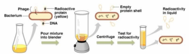
The simple viral-host system of T2 bacteriophage and E. coli was the perfect battleground on which to pit the protein theory of heredity against its DNA competitor. The reason being that T2 phage is made up of only two parts: a protein coat and the DNA contained therein.

Just months before the blender experiment, electron micrographs taken by Thomas Anderson showed that phage never invades bacteria as a whole: it always remains attached at the surface. Some part of the phage – either its DNA or certain proteins – must enter the bacterium in order to hijack its cellular machinery and produce new virus.

In an experiment many biologists would gladly give their pipetting arm to have designed, Hershey and Chase tracked the location of T2 phage DNA during its hostile takeover of E. coli by tagging it with a radioactive form of phosphorous. DNA contains vastly greater amounts of phosphorous compared to protein, so the presence of high levels of radiation would give away phage DNA’s location. Hershey and Chase wanted the tagged phage to infect bacteria, but they didn’t want it to stick around at the surface – they only cared about what the phage transferred to the bacterium during the initial stage of infection.

This is where the Waring blender got its chance to shine.

The blender wasn’t their first choice for removing phage from the outside of bacteria. According to Hershey, “We tried various grinding arrangements, with results that weren’t very encouraging”. It was only after a fellow CSHL geneticist, Margaret McDonald, offered her blender that the experiments started to work. The Waring blender had just the right amount of shearing force to remove phage coats from the bacterial walls, without rupturing the bacteria completely. They then spun the bacteria/phage smoothie in a centrifuge to separate infected bacteria from everything else: bacteria formed a pellet at the bottom of the tube, while everything that was smaller (including protein coats) stayed in the liquid above it. Upon measuring radioactive phosphorous, Hershey and Chase found that only bacteria showed high levels of radiation. DNA made it into the bacteria, protein didn’t. Most importantly, phage were able to replicate in bacteria even when their protein coats were kicked off soon after infection.

DNAlabellingchase

It looked like protein itself wasn’t necessary for phage reproduction after all. It might just serve as a passive vehicle to get the genetic material into the cell. At first, Hershey thought there must be a mistake. After presenting the blender experiment results in a staff meeting, a young scientist named Waclaw Szybalski recalled Hershey said, “I don’t believe in that DNA”. Meanwhile, Barbara McClintock thought it was a nice experiment.

Hershey’s skepticism is a testament to his scientific rigor, considering that he stared at a near perfect explanation for his results around this time. In a letter to Hershey, Roger Herriott wrote,

I’ve been thinking – and perhaps you have been too – that the virus may act like a little hypodermic needle full of transforming principles; that only the tail [of the virus] contacts the host and perhaps [sic] cuts a small hole through the outer membrane and then the nucleic acid of the virus head flows into the cell.

Hershey and Chase repeated their experiment, only this time they tagged the phage protein by radiolabeling an element found only in protein: sulfur. They found the exact opposite result: radiation was found in the liquid portion that contained the protein coats and not in the bacteria. Again, the virus was able to replicate in the bacteria cells after blending had removed protein coats from the cell walls. Further experiments showed that phage offspring contain radioactive phosphorous passed down from the tagged parental phage. Virtually no radioactive phosphorous is found in the protein of viral replicates. This finding further solidified the notion that DNA was the genetic material responsible for reproduction.

proteinlabelchase

A mere 11 months after reading a letter from Hershey detailing the results, James Watson along with Francis Crick solved DNA’s structure, unlocking its hidden code. The Hershey-Chase experiment convinced Watson that the 3D structure of DNA was worth chasing, and to say that DNA was the next big thing in biology would be an understatement. Still, Hershey’s cautious personality shone through the last sentence of his and Chase’s groundbreaking paper, “Further chemical inferences should not be drawn from the experiments presented.”

*******

This post is a result of a CSHL tour I gave to a high school class this week. The teacher asked me in which building Hershey and Chase performed their blender experiment. I took a safe guess at McClintock Laboratory, which back in the day was the animal house. While correct, it made me want to refresh my memory of the experiment. As a working scientist, I have a much greater appreciation for its simplicity and elegance than I could at 15.

Martha Chase and Alfred Hershey at CSHL, 1953

Just as I naively took “Waring blender” to be a single entity, I saw an equal scientific partnership in the symmetry of the “Hershey-Chase” experiment’s name. Instead, Martha Chase was Hershey’s young lab technician. Hershey wrote that the two were “groping toward the idea of the blender experiment” in 1951, implying that she did aid in the experiment’s design. However, I have yet to find any account of the work in Martha’s own words. While Hershey went on to win the Nobel Prize in 1969 with other Phage Group pioneers, Max Delbruck and Salvador Luria, Martha’s life took a more tragic trajectory.

In my reading I came across a post on a blog about the scientist Linus Pauling called “The Martha Chase Effect.” The author found that while their blog was dedicated to the life and work of Linus Pauling, the search term that most frequently brought people to the site was, “Martha Chase”. This is because Chase’s digital footprint, particularly for images, “represents a much smaller body of water” than Pauling’s.

A puddle, really.

In a future post, I hope to explore the “Martha Chase Effect” and learn more about the young woman whose name is forever eponymous with the Waring blender experiment.

Edit:

Take a gander at the actual blender used in the experiment: not as sleek as the one I showed  (notice the warning label for radioactive P32).

Thanks to Matthew Cobb for the link!

Sources:

Thanks to The Linus Pauling blog, for background information and inspiration for the Martha Chase Effect.

“A Phage Shows its Claws” by Lee Simon in New Scientist and Science Journal, 1971.

“The Injection of DNA into Cells by Phage” by A.D. Hershey from Phage and the Origin of Molecular Biology CSHL Press.

Martha Chase Dies” by Milly Dawson in The Scientist Aug 20, 2003.

“Independent Functions of Viral Protein and Nucleic Acid in Growth of Bacteriophage” by Hershey and Chase in The Journal of General Physiology. 1952

CSHL Oral Histories: Waclaw Szybalski on Martha Chase.

Carl Zimmer’s advice for aspiring science writers

It’s a daunting time to be a professional science writer. Science, it seems, is working too well. As Carl Zimmer told an auditorium full of science graduate students, “It’s hopeless to cover it all, and it’s only getting worse.”

Or as an exasperated Ed Yong put it:

edyong

This point in the history of science represents an embarrassment of riches – exciting discoveries are made every day, but there aren’t enough people to take scientific reports and craft them into stories that non-scientists understand and actually want to read. As Zimmer told those of us gathered in the auditorium of Yale’s Peabody Natural History Museum, “Let’s not restrict the wealth to this room.”

There is, in fact, a world outside of our departments, and there’s a lot at stake out there.

Nearly half of Americans believe that humans were created in their present form by God within the past 10,000 years. The percentage of Americans who hold a hard-line Creationist view of the world has been flat for the past 30 years, despite major the gains science has made in understanding our human origin, including sequencing of the human and chimp genomes. The mounting scientific evidence that humans and chimps evolved from a common ancestor ~5-7 million years ago has failed to make those data points budge. Consequently, Americans still debate what place human evolution has in the classroom. Similarly, evidence of a 150-year trend in rising global temperatures hasn’t stopped people from pointing to a cold winter’s day as a refutation of climate change.

gallupevo

The public policy that will determine what we teach our kids and how we deal with climate change will hinge on whether or not voters understand the scientific method, or at least value scientific evidence. Scientists can’t afford to discuss amongst themselves and grumble about how the public doesn’t get it. Fortunately, if the attendance at Carl Zimmer’s writing workshop is any indication, scientists are looking to add their voices to the public discourse. If not to increase science literacy then, heck, to at least share the exciting things happening in their labs with family and friends. Scientists can chip in to cover “all the amazing papers”, lest Ed Yong sends himself to an early grave.

So great, we have scientists who are onboard and want to deliver science to the public. One problem: scientists, we’re told, are pretty bad at communicating science.

Take this tweet from Quartz News tech/science journalist Christopher Mims, posted that very same afternoon:

mimsOuch.

Or consider this quote about scientists-as-writers from Robert Kunzig from “Gee Whiz Science Writing” in A Field Guide for Science Writers:

It was sometimes almost sweet, how incompetent they were- how unable to offer a clear, logical account of their work that would be understandable and interesting to an intelligent layperson.

I read this passage on the eve of the writing workshop. It fell under the subheader “Don’t Be Afraid” and it was aimed at convincing science journalists that they shouldn’t be intimidated by egg-headed scientists. As an egg-headed scientist it left me feeling, well, intimidated at the prospect of a professional writer ever reading my own attempts at science writing.

But here was acclaimed science writer Carl Zimmer telling us that we too could learn to be effective writers. It would just require a bit of “deprogramming”. The thing is, scientists sometimes forget to speak English. As students we put in a lot of effort to learn the scientific language, or jargon, of our fields – terms like “synaptic plasticity” or “epigenetics” encapsulate a textbook worth of material. We embraced jargon because we earned the right to use it, and as Zimmer said it, we wanted to “sound like the grownups”.

Jargon is super convenient when experts wish to convey concepts quickly among themselves, but it’s also a sure-fire way to make the eyes of non-scientists glaze over faster than you can say “Krispy Kreme”. First and foremost, scientists need to dispense with scientific lingo when they write for the public. Some jargon is easy to spot, like “RNA interference”. However, scientists sometimes have “jargony” uses for everyday English words that are confusing to lay readers. I thought this table from “Communicating the science of climate change” by Richard Somervile and Susan Hassol was an eye-opener.

sciterms

Instead of jargon, scientists should find metaphors that bring concepts to life and help readers get an intuitive sense of the science. A spot-on metaphor or figure of speech doesn’t exclude readers, but like jargon it quickly conveys a complex concept. Zimmer pointed out, “our brains are built for metaphors”. To avoid “in-speak” scientists need to place themselves in the mind of a non-scientist, which requires a forgetting of sorts. Think: what did words mean before science colored them? Zimmer keeps a running log of banned words in science writing that you can refer to. I’m guilty of relying on “forbidden” words like modulate and elucidate, among others.

Zimmer said he’s noticed that jargon is leaky, often seeping out into surrounding sentences. That is, scientists tend to write in overly formal language. A real-life example he used was the graduate student who wrote that flu spreads in “household settings”. Another bad habit of scientists is that they often use the passive voice. I think this happens for two reasons: 1) scientists are typically modest and sometimes feel like saying I or we did/found X sounds arrogant 2) scientists aim to be unbiased; when the passive voice is used in scientific articles it’s as if to say “any person who followed the stated protocol would have found the same results”. However, when passive voice creeps into non-academic writing, it can leave the reader with the impression that phenomena unfolded without any apparent cause.

Once a scientist unlearns his or her bad habits, the next step is to practice the craft of story building. Zimmer said that when we look at a beautiful piece of architecture, what we appreciate is how the craftsmanship at different scales work together. In writing, the craft starts with word selection and scales up to the paragraphs and overall structure of the story. Like a building, we can take apart the elements of a story and figure out how it was constructed and what makes it compelling. If you want to learn how to write a magazine article, reverse engineer a magazine article.

Zimmer said that before starting, you should be able to state your story in ten words or less. If you can’t, you don’t know what you’re writing about yet. The introduction should provide a road map for the journey you’ll take the reader on. The end of the story can look towards the future, “What are the implications if this finding holds up?” Stories, scientific ones included, are about people doing things – so bring the players to life. Also, writers should make sure to orient the reader throughout the story, especially if the piece jumps around in time. Finally, don’t be afraid to read your writing out loud. The most enjoyable writing flows naturally and takes on the cadence of speech.

Scientific trainees spend much more time wielding pipettes than wielding pens. Yet, the ability to write clearly for a lay audience is vital if scientists want to inform public opinion on issues ranging from food production to climate change. At a time of looming budget cuts, scientists need the support of the public. We should be seeking to communicate how our science can improve lives, whether that means finding new energy sources or simply providing people with a “gee whiz” science moment. We might need to unlearn some habits in order to do it, but scientists can help spread the wealth of science to the public.

Zimmer’s workshop laid out specific tools and guidelines to start the process, but it will take a lot of work from there. Luckily, as scientists we already possess something that’s key to good story-telling, and that’s enthusiasm. Former science editor for The Guardian Tim Radford wrote:

Most scientists start with the engaging quality of enthusiasm — to get through a degree course, the PhD and all the research-council hoops, you would need it…Enthusiasm is infectious, but to command an audience of readers, scientists should exploit their other natural gifts. One of these is training in clarity. Another is training in observation. And a third is knowledge.

I’ll keep these words in mind as I work to put Zimmer’s writing advice into practice.

Why a promising experimental result is like a new boyfriend.

There may have been others before you, but I can you tell this time is different. You won’t deceive me, and what I’ve found in you will be true. You won’t be like the others, who raised my hopes and had me planning for a future together. A future that was never realized. The follow-up experiments that were never performed, the papers that went unwritten.

And I admit it. It was my fault.

Prematurely, I placed significance on relationships that were new and uncertain, that hadn’t yet been tried and tested. I did this because I wanted so badly for each one to work out. My heart swelled at the slightest suggestion of a correspondence between my deepest hypotheses and desires and my experimental results. I wanted to find meaning in it all, to know that all of the time and effort I had invested in the lab wasn’t in vain. That my work would lead me to that feeling of discovery that happens when you find the right one. The one who opens your eyes to new possibilities and forever changes the course of your thesis project.

But I’m not a first year graduate student anymore. I guess you can say I’ve built up my defenses. I don’t fall so easily. They say that love is blind, and you know what – it’s probably best that the experimenter is too.

Still, that didn’t stop me from getting butterflies the first time I saw you. But this time I kept my cool. I took my time getting to know you. I checked that you were consistent, saw how you behaved in different conditions. I made sure that you’d stick around before I ran to tell my labmates about you. And now I can say that I think I’ve found the real deal. This relationship looks like it’s too significant to fail.

My n is high and my p value is low, and this time, baby, I think we’re going to make it.

Move over Martinotti cells.

GABAergic inhibitory interneurons (INs) account for approximately 10-20% of neurons in the rodent cortex. Of these, about 20-30% express a peptide called somatostatin (SOM). Most study into the function of SOM INs has focused on one class in particular called the Martinotti cells. Martinotti cells have axons that ascend to the most superficial layer of the cortex, contacting distal sites on the dendrites of excitatory pyramidal cells. Recent papers, like this one from Massimo Scanziani’s group, highlight the role of layer 2/3 SOM INs in mediating surround suppression of layer 2/3 pyramidal neurons. Feedback inhibition from SOM INs is recruited via excitatory input coming from the horizontal axons of pyramidal neurons. In this way, inhibition from SOM INs scales with the spread of excitation in layer 2/3.

Given what was known about SOM INs, researchers in Bernardo Rudy’s group at NYU were surprised to find that when they optogenetically silenced the activity of SOM INs in brain slices (using conditionally-expressed halorhodpsin in the SOM-Cre mouse line), layer 4 pyramidal neurons fired less. Meanwhile, layer 2/3 pyramidal neurons fired more, as one would expect upon removing a source of inhibition.

The key to this result lies in the distinct pattern of connections that layer 4 SOM INs exhibit compared to their Martinotti cousins.

L: SOM-expressing Martinotti IN on the left displays typical axonal morphology that extends into layer 1. R: a layer 4 SOM IN with an axonal arbor that is restricted to layer 4.

Left: Martinotti IN displays ascending layer 1 axons. Right: axonal arbor of layer 4 SOM IN is restricted to layer 4

Analysis of layer 4 SOM INs revealed that their axonal arbors are restricted to layer 4, the layer of the cortex that receives thalamic input. In addition, layer 4 SOM INs exhibit electrophysiological properties that are distinct from Martinotti cells: notably, they have a more hyperpolarized resting potential and can fire at much higher rates. What about their functional connectivity?

Dual recordings between SOM and either pyramidal neurons or fast-spiking (FS) GABAergic INs revealed that SOM INs were much more likely to be connected to FS INs than pyramidal neurons. The connection probability from SOM INs to FS INs in layer 4 was surprisingly high: 62%. Furthermore, measures of unitary IPSCs and IPSPs in response to SOM IN spiking revealed that response amplitude was consistently higher in FS INs compared to pyramidal neurons. This finding was also (rather heroically) confirmed by exciting a SOM IN and simultaneously recording responses in a FS IN and pyramidal neuron. Keep in mind that only SOM INs were fluorescently-tagged, so that FS INs had to be hunted for the old-fashioned way – by taking a guess, patching a selected neuron, and testing its spike properties.

FS INs show larger inhibitory responses to SOM IN activation in layer 4, while pyramidal neurons show greater responses in layer 2/3.

FS INs show larger inhibitory responses to SOM IN activation in layer 4, while pyramidal neurons show greater response in layer 2/3.

Importantly, short term dynamics were similar across the two types of synapses. Unitary IPSCs evoked by SOM INs were moderately depressing in both FS INs and pyramidal neuron synapses confirming that FS INs really do “see” more inhibition than pyramidal neurons, whether SOM INs fire sparsely or repetitively. When the same experiments were performed in layer 2/3 the results were flipped: SOM INs were more likely to contact pyramidal neurons, and they inhibited them more strongly than FS INs. This set of experiments was repeated using a sexier approach (*cough* optogenetics) that produced the same results. Experimenters conditionally expressed ChR2 in SOM INs by injecting a virus encoding floxed ChR2 into the SOM Cre mouse line. They then recorded the light-evoked IPSCs in FS INs and pyramidal neurons.

Given that the major role of layer 4 SOM INs, as defined by their functional connectivity, is to inhibit FS INs, it makes sense that silencing SOM INs reduces pyramidal neuron firing in layer 4. Based on connectivity, one would predict that silencing layer 4 SOM INs would remove inhibition placed on FS INs. Once disinhibited, FS INs would fire more and inhibit pyramidal neurons to a greater extent.

Yellow bar indicates SOM IN inactivation. FS INs fire more  when SOM IN silent while pyramidal neurons (PN) fire less

Yellow bars indicate SOM IN inactivation. Arrows indicate thalamic stimulation. Layer 4 FS INs fire more when SOM INs are silenced while pyramidal neurons (PN) fire less.

Indeed, researchers showed that optogenetically silencing SOM INs increased the number of spikes that layer 4 FS IN s fired during UP-states. Finally, to demonstrate that FS INs are the potent inhibitors of layer 4 pyramidal neurons scientists silenced FS INs using halorhodopsin expressed conditionally in the PV-Cre mouse line. Inhibiting PV INs significantly increased pyramidal neuron firing, supporting the notion that layer 4 SOM INs could indirectly excite pyramidal neurons by inhibiting PV fast-spiking interneurons.

Most studies on inhibitory neuronal circuits have focused on the control that GABAergic interneurons can exert over excitatory neuronal activity, but we are beginning to uncover the interactions among GABAergic interneurons. These interactions may potentially form disinhibitory circuits that could serve to gate the transmission of information to excitatory pyramidal neurons. Andres Luthi’s lab reported the existence of a disinhibitory microcircuit that facilitates learning of an associative fear memory. Foot shock excites layer 1 INs in auditory cortex that in turn inhibit FS INs in layer 2/3. This results in disinhibition of pyramidal neurons that was necessary for acquisition of the auditory fear memory.

Because all of the experiments regarding layer 4 SOM INs were performed in brain slices, we don’t know when this disinhibitory circuit could be activated in the intact brain. Previous studies suggest that layer 4 SOM INs only receive weak thalamocortical input, so it is unlikely that this disinhibitory circuit is recruited directly by thalamic activity. However, layer 4 SOM INs are potently activated by cholinergic inputs. An appealing possibility is that during heightened states of arousal cholinergic inputs excite layer 4 SOM INs. In turn, layer 4 SOM INs inhibit FS INs, thereby facilitating transmission of sensory information from the thalamus to the cortex. This study raises interesting questions to be addressed in vivo.

*EDIT* thanks to Nick Olivas who reminded me of this 2012 paper studying the function of SOM INs in vivo during active whisking. It’s interesting to note that researchers found that layer 2/3 SOM INs become hyperpolarized during active whisking, increasing the excitability of pyramidal neuron dendrites. If at the same time layer 4 SOM INs are excited, layer 2/3 pyramidal firing could be driven further by increasing the excitatory relay from layer 4 pyramidal neurons onto readily-excitable layer 2/3 neurons.

“Just keep swimming”- illuminating circuits of motivation

ImageNeuroscientists at Stanford have discovered a brain pathway that controls a rat’s will to fight in a challenging situation.

You’ve probably seen the “Hang in there!” motivational poster – the rat version would feature a tank of water with the caption,”Just keep swimming!” For decades, scientists have used the forced swim test (FST) as a means to model depressive behavior in rodents. This simple test measures what a rat does when it’s placed in a tank of water that it can’t escape – does it keep still and float or does it fight like hell to get out?

Just as people handle stress differently, rats display a range of responses in the FST. Researchers quantify the time spent actively trying to escape (i.e. kicking and climbing) versus time spent passively floating, with the idea that rats that are behaviorally depressed give up more easily whereas resilient rats keep kicking. Immobility during the FST is interpreted as behavioral despair or “checking out” as a means of coping with stress. The switch from fighting to floating may reflect a rat’s assessment that kicking is probably useless. Similarly in depression, pessimism regarding one’s control over a situation can lead to feelings of hopelessness and a loss of motivation. Further evidence that the FST relates to human depression is the ability of factors like antidepressants and stress to alter the amount of time rodents spend immobile. In fact, FST is the most widely used behavior to test efficacy of antidepressants in lab rodents, with reduced immobility indicating a positive drug effect. You can check out a video of the behavior here.

ImageScientists in Karl Deisseroth‘s lab wanted to identify a brain circuit that determines whether a rat selects an effortful or inactive behavior when challenged. In theory, FST was an ideal behavior because the transitions from active escape behavior to passive floating are clearly demarcated. Typically, FST activity is measured from video footage by motion detection software and human raters. However, this level of detail wouldn’t cut it for Dr. Melissa Warden and colleagues, who wanted to link real-time changes in brain activity to behavior. They came up with a simple yet ingenious hack to generate precise, time-stamped activity data: by placing an induction coil around the tank and attaching a small magnet the rat’s hind paw, a small current was generated each time the rat kicked.

In search of an action selection circuit, Warden and colleagues recorded neuronal activity in the prefrontal cortex (PFC), a region that’s important for coordinating thoughts and behavior. The PFC’s ability to select appropriate actions to match internal goals is no simple task: an animal has to maintain a goal in mind (say, “mate”), plan actions (approach the female), and update its strategy based on feedback (she is not into it). Several notable human cases illustrate the PFC’s complicated role in action selection. Take Phineas Gage, whose personality was dramatically altered after he took an iron tamping rod through the PFC. It seemed that the once polite man lost his “filter” and was unable to resist the urge to make inappropriate remarks and curse like a sailor. On the flip side, some recipients of the infamous prefrontal lobotomy – a procedure that intentionally damaged the PFC – experienced a severe loss of will and motivation. The fact that PFC damage can affect motivational state in opposite directions suggested to researchers that they’d find a mix of neuronal responses in the PFC during the FST, with no simple “on means kick” type of phenomenon.

Arrays of individual PFC neurons were recorded before, during, and after the FST. Researchers predicted that activity of action selection neurons would be modulated by behavioral state within the context of the FST. Many neurons showed a straight-forward correspondence between firing rate and motion, most-likely reflecting motor activity and not behavioral control. However, a subset of neurons showed a surprising activity profile: they were highly active during the largely immobile pre- and post- FST periods, but during the FST they were active during kicking and silent during floating. Other neurons were more active during the mobile phase of the FST and appeared to turn on before kicks. Overall, the activity profiles were a mixed-bag, but researchers identified subsets of neurons that were shut-off during floating or that turned-on to “kick-start” escape behavior.

Turning up the volume on an entire party would drown out the message of a small group.

Despite mixed neuronal responses, researchers went ahead and tested if activating PFC neurons during the FST changed mobility. They injected a virus encoding instructions for making channel-rhodopsin (ChR2) into the PFC of rats. When ChR2 is expressed in neurons, blue light causes the channel to open, allowing positive ions to flow in and drive neuronal firing. When Warden and colleagues illuminated ChR2-expressing PFC neurons in the FST they saw no change in mobility. This is not so surprising, given how varied the neuronal activity profiles were: if only a small subset of neurons carried the signal for “SWIM!”, the signal would be lost in the noise when all PFC neurons were activated. Imagine a small group of people scattered throughout a crowded cocktail party who are all saying the same thing. If the volume were turned up on the entire party, this message would be drowned out by chatter. The researchers now needed to narrow their manipulation to a select group of neurons that could influence behavior in the FST.

Warden and colleagues illuminated the DRN to selectively activate input from PFC neurons that expressed ChR2.

Warden and colleagues illuminated ChR2 positive terminals in the DRN to selectively activate input from PFC.

Warden and colleagues turned their attention to the dorsal raphe nucleus (DRN), a region that the PFC projects to that has also been implicated in motivated behavior and depression. Selectively activating the PFC–>DRN projection was a simple matter of aiming the light. ChR2 is not only expressed on cell bodies but can also be found at the axon terminal, the site of neuron-to-neuron communication. ChR2 virus was injected into the PFC, and the DRN was illuminated, activating input from the PFC. This time, the results were dramatic.

When the light was switched on-and-off for 2 minute periods, kick frequency rose and fell with the “flip” of the light switch. Importantly, activating the PFC input didn’t increase locomotion outside of the FST, suggesting PFC–>DRN pathway activation specifically drives effortful behavior when the animal is challenged. In addition, pharmacologically blocking excitatory transmission at the PFC–>DRN synapse eliminated the effect. Interestingly, when they injected ChR2 virus into DRN and directly activated it, they found mobility was increased both in and out of the context of the FST. These differences in behavior suggest that PFC targets a specific subset of DRN neurons. Finally, when they performed the same experiment in the lateral habenula (yep, another brain area implicated in depression) they observed the opposite effect, with activation leading to a rapid reduction in mobility.

kickfreq

Kick on, kick off. Light activation of the PFC to DRN projection increases kick frequency during the FST. Turn off the light and frequency drops.

This study sends the message that in the brain, it’s not just about “who’s talking” but “who’s talking to whom”. This is clearly demonstrated by the observation that activating a mixed population of neurons in the PFC has no effect on behavior, while activating PFC axons at their target sites has profound and diverse effects. This study also highlights how the field of neuroscience has moved beyond the idea that “brain area x does y” and recognizes that it takes distributed circuits of neurons to carry out complex behaviors. With the wide variety of optogenetic tools available, we’re in a heyday of this type of circuits-to-behavior study. This paper is noteworthy for its creative solutions to studying the neural mechanisms underlying the widely-used FST and for the identification of divergent PFC pathways that alter the rat’s will to fight under challenging conditions. Finally, the ability of PFC projections to DRN and LHb to alter motivational state makes predictions for how these pathways may be dysregulated in human depression.

(Shop-talk below)

Did this paper accomplish what it set out to do, to find the PFC circuitry that determines whether effortful or inactive behavior is selected in challenging situations? The ability of the PFC–>DRN pathway to initiate escape related behaviors and PFC–>LHb to switch kicking to floating was quite dramatic. However, there is a question of how artificial activation of this pathway relates to the normal FST behavior. Initial recordings in PFC identified a subset of neurons that were modulated by FST in a way that could reflect action selection. However, the authors skipped ahead to (somewhat arbitrarily) activate the PFC–>DRN pathway. It’s implied, but never demonstrated, that the neurons that are selectively silenced during the immobile phase of the FST are the DRN-projecting neurons. This experiment could be done by using a 2 virus system that requires retrograde infection of PFC neurons from DRN plus local viral injection in PFC in order to express ChR2. PFC neurons could be chronically recorded across pre-FST, FST, and post-FST and the DRN-projecting neurons identified via the PINP method.

Resources:

Warden M, Selimbeyoglu A, Mirzabekov J, Lo M, Thompson K, Kim S, Adhikari A, Tye K, Frank L, and Deisseroth K. A prefrontal cortex–brainstem neuronal projection that controls response to behavioural challenge. Nature. 18 Nov. 2012.

Slattery D and Cryan J. Using the rat forced swim test to assess antidepressant-like activity in rodents. Nature protocols. 3 May 2012.

Like son like…mother? Macro problems with a recent paper on microchimerism

Image

Gene found on Y chromosome detected in females brains.

A recent Plos One article found expression of a male-specific gene in a high proportion of female brains, providing the first evidence for microchimerism in the human brain (it’s been shown in mice). Furthermore, researchers reported that the brains of women who had Alzheimer’s disease were less likely to harbor male DNA.

Microchimerism refers to the persistence of a small number of genetically distinct cells – or just the DNA – within a host. During pregnancy, cells are exchanged between mother and fetus via the placenta and can reside in a variety of organs for the lifespans of mother and child. Studies have established that microchimerism can have either detrimental or beneficial effects for mother and offspring, as in the case of autoimmunity or cancer, respectively. Imagine the fascinating implications of a child’s cells living in his mother’s brain.

Back up a minute. Before we’re overcome with the warm and fuzzies thinking about nature’s nod to the amazing bond between mother and child, let’s first consider the primary article in question. One glaring problem: we don’t know how many of the women were, in fact, mothers.

Pregnancy history was largely unavailable to the researchers, but let’s look at the cases for which they knew maternal status: in 5/9 cases of women who had had sons, male DNA was detected. In 1 female who had no history of having a son, male DNA was detected. To be fair, the presence of the male DNA could reveal an unknown miscarriage. However, male DNA can also be transferred via mechanisms that are not dependent on maternal status, such as from a twin brother or even an older brother who previously took up residence in his mother’s uterus and made her microchimeric. This limitation is an even bigger problem when the researchers seek to compare brains of control females and those with Alzheimer’s disease (AD) – what if mothers were unequally distributed across groups? If maternal history were available, it would have been interesting to know if there was a dose-dependent effect (more sons = greater concentration of male DNA?) or how failed pregnancies may contribute to microchimerism compared to successful pregnancies.

Because Alzheimer’s disease (AD) has been reported to be more prevalent in mothers than women who do not have children, researchers compared male DNA levels in neurologically normal women and AD patients. I mentioned the potential complication that the control group may have included more mothers, but beyond that there are flaws in the study’s design. Postmortem brain studies face many challenges, particularly when scientists wish to apply molecular techniques to the tissue. After a patient dies, it’s a race against the clock to section the brain and immerse it in fixative. The so-called post mortem interval (PMI) is a crucial factor for preserving the integrity of proteins and genetic material. To deal with the inevitable uncontrolled variables that pop up during tissue processing, researchers try to get all their ducks-in-a-row by matching subjects beforehand. This means that one would compare a 70-year-old woman with AD whose brain was fixed after 18 hours with a woman of a similar age and PMI. While the researchers in this study used statistical methods to correct for subject differences, the subjects were not matched, and the AD group was significantly older. Therefore, tissue quality could be reduced in the AD group, making it more difficult to detect the male genetic material.

Despite drawbacks, it is still striking that 18/26 control female brains had detectable levels of the male gene in at least one brain section of brain sampled – suggesting that microchimerism is prevalent. However, the case is not strong that there is any difference between control and AD patients. When the researchers analyzed the concentration of male microchimerism they did not find a statistical difference after excluding an outlier in the control group who had 10 times more male DNA than the next highest sample (did she have a brood of boys?!). They also had a wonky design whereby they measured male DNA in whatever brain samples were available – a bit of hippocampus, a little pons here.. without regard for comparing similar samples across groups. Therefore, when they report the proportion of positive samples across all regions, it isn’t a fair comparison. Add in some uncertainty as to how many mothers were in each group, and it seems that any conclusion regarding differences between AD and control subjects is shaky at best.

I could let my imagination run wild with follow-up questions like, “Are there fewer positive samples in AD patients because chimeric cells are more vulnerable to AD-induced cell death?” but these findings first require further validation in patients whose maternal status is documented. Still, studies like this that suggest widespread microchimerism open up fascinating avenues of research into the role that these “immigrant” cells play in normal health.

*EDIT: I wanted to point out that chimerism isn’t unique to males. This study chose to look at the Y chromosome because females are XX, so it would be easy to detect chimerism due to the fact that there shouldn’t be any extra Y chromosomes hanging around.